Please wait a minute...
Welcome to Bulletin of Botanical Research! Today is Share:
IMAGE/TABLE DETAILS
In vitro Germination,Callus Induction and Primary Haustorium Organogenesis in the Parasitic Plant Cynomorium songaricum
Xin YUE, Gui-Lin CHEN
Bulletin of Botanical Research    2020, 40 (6): 846-854.   DOI: 10.7525/j.issn.1673-5102.2020.06.007
Abstract   (657 HTML13 PDF(pc) (3055KB)(271)  

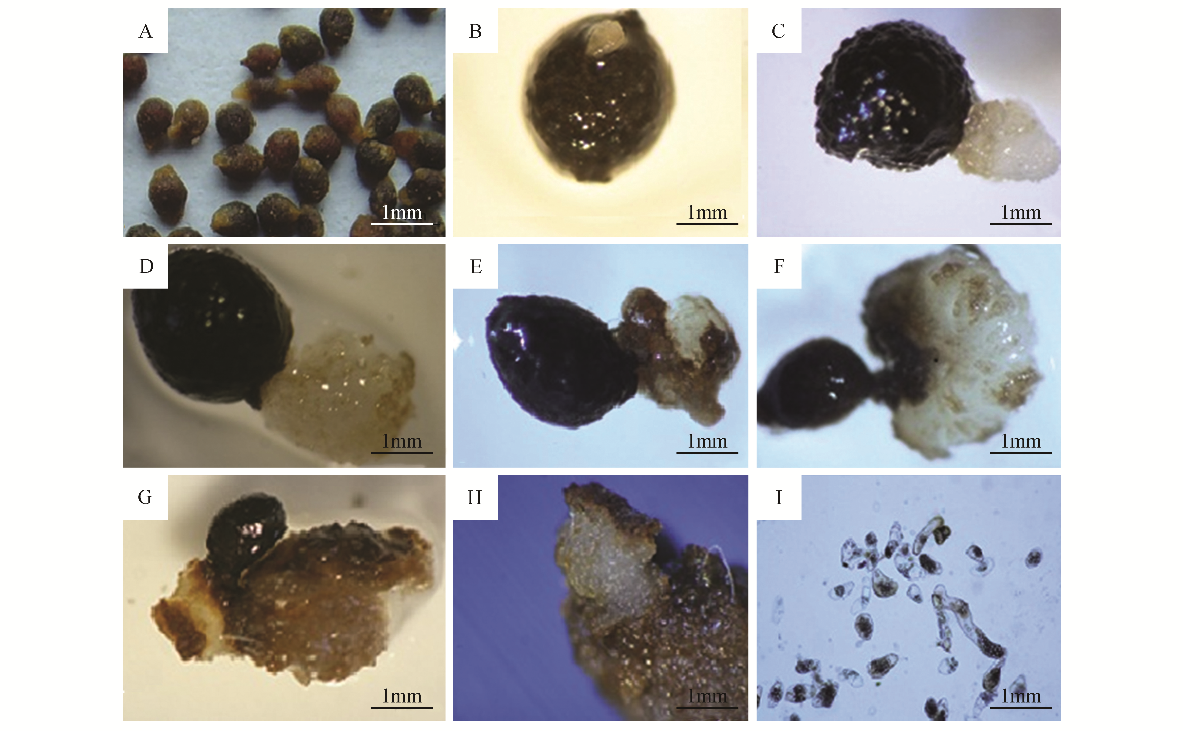
Cynomorium songaricum Rupr. is an obligate parasitic plant widely used as traditional Chinese medicine and Mongolian medicine. Here, we firstly describe protocols for in vitro germination, callus induction and haustorium organogenesis in C.songaricum Rupr. In this study, adequate concentrations of gibberellic acid(GA3),in combination with other plant growth regulators in the medium, promoted embryo development and germination of C.songaricum seeds. The highest callus induction rate from seeds(13.7%) after a 40 d incubation was obtained with B5 medium adding 2,4-dichlorophenoxyacetic acid(2,4-D; 1.0 mg·L-1), kinetin(KT;0.5 mg·L-1) and GA3(1.0 mg·L-1. This resulted in callusformation in 13.7%±3.1% of seeds. Addition of 2,4-D(0.5 mg·L-1) and KT(0.25 mg·L-1) yielded highest haustorium organogenesis from calluses. Some primary haustorium branched to form adventitious roots of 3-4 cm in length. Subsequently, the tip of each adventitious root formed a nascent primary haustorium, which was then branched out into adventitious roots. The role of auxins(2,4-D in this study) in the formation of primary haustorium and adventitious roots from seed callus in C.songaricum was also discussed.


Fig.2 Callus development in different periods of cultivation in C.songaricum
The embryo broke through the seed coat at the micropylar end after 40 days of culture(Fig.2B). The radicle continued to elongate in the following days(Fig.2C). The radicle’s top became intumescent(Fig.2D) and eventually formed a callus(Fig.2E). The callus was pure white and dense, and grew rapidly from the radicle(Fig.2F). After another 20 d, it became brown(Fig.2G), and embryogenic callus formation occurred(Fig.2H). Embryogenic cells were mostly cylindrical, with large nuclei and starch grains(Fig.2I)
Extracts from the Article
The embryo broke through the seed coat at the micropylar end after 40 d culture(Fig.2B), and the radicle continued to elongate in subsequent days(Fig.2C). The radicle’s top became intumescent(Fig.2D) and eventually formed the callus(Fig.2E), which was pure white and dense, growing rapidly from the radicle(Fig.2F). After another 20 d, it turned brown(Fig.2G), and embryogenic callus formation occurred(Fig.2H). Embryogenic cells were mostly cylindrical, with large nuclei and starch grains(Fig.2I). The rates of callus formation from C.songaricum seeds were assessed in the dark under various combinations of three hormonal plant growth regulators(Table 1). The highest callus induction rate(13.7%) after a 40 d incubation was produced by the combination of 1, 0.5 and 1 mg·mL-1 of 2,4-D, KT, and GA3, respectively. This rate was significantly higher than those obtained with other combinations.
The embryo broke through the seed coat at the micropylar end after 40 days of culture(Fig.2B). The radicle continued to elongate in the following days(Fig.2C). The radicle’s top became intumescent(Fig.2D) and eventually formed a callus(Fig.2E). The callus was pure white and dense, and grew rapidly from the radicle(Fig.2F). After another 20 d, it became brown(Fig.2G), and embryogenic callus formation occurred(Fig.2H). Embryogenic cells were mostly cylindrical, with large nuclei and starch grains(Fig.2I)
Other Images/Table from this Article